Art-labeling activity anatomy and histology of the adrenal gland – Embark on an art-labeling adventure into the realm of anatomy and histology as we delve into the intricate details of the adrenal gland. This engaging activity invites you to become an artistic explorer, meticulously labeling the histological slides of this enigmatic organ, uncovering its hidden secrets and gaining a profound understanding of its structure and function.
Prepare to embark on a journey of discovery, where each brushstroke unveils a new layer of knowledge, transforming you into a master of adrenal gland anatomy and histology.
Art-Labeling Activity: Anatomy and Histology of the Adrenal Gland: Art-labeling Activity Anatomy And Histology Of The Adrenal Gland

Introduction
Art-labeling activities are a valuable tool in anatomy and histology education, allowing students to engage actively with the subject matter and enhance their understanding of complex structures. This activity focuses on the anatomy and histology of the adrenal gland, an essential component of the endocrine system.
The adrenal gland plays a crucial role in regulating the body’s response to stress and maintaining homeostasis. By understanding its anatomical and histological features, students gain insights into its function and clinical significance.
Materials
- Histological slides of the adrenal gland
- Microscope
- Labeling tools (e.g., colored pencils, markers)
- Reference materials (e.g., textbook, atlas)
Preparation of Histological Slides
1. Obtain histological slides of the adrenal gland, ensuring they are properly stained and mounted.
2. Place the slides under a microscope and adjust the magnification to observe the tissue.
Procedure
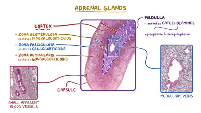
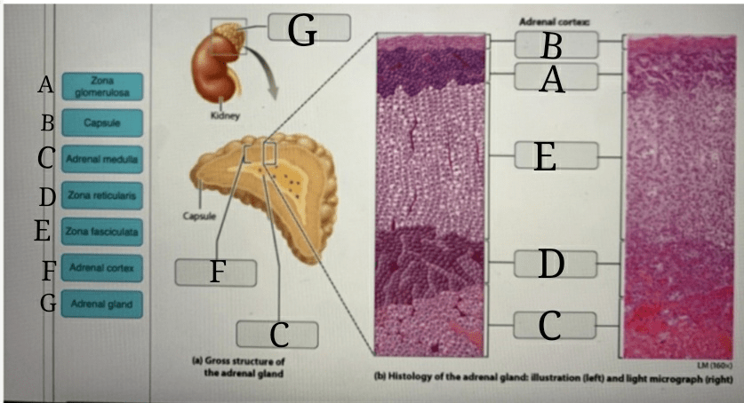
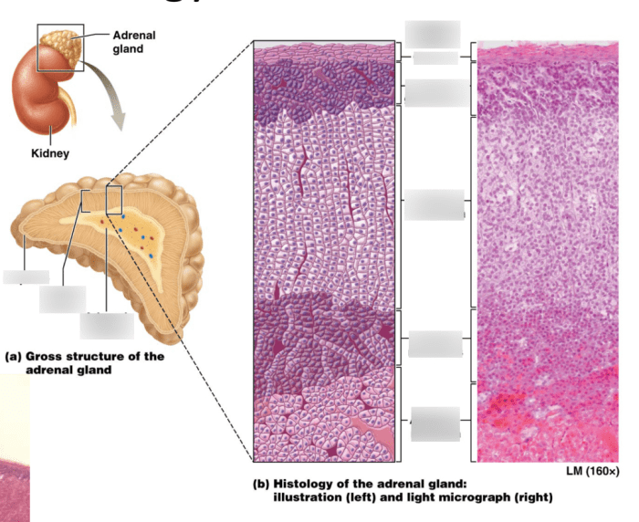
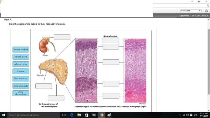
- Identify the different zones of the adrenal gland: the zona glomerulosa, zona fasciculata, and zona reticularis.
- Label the adrenal cortex, adrenal medulla, and capsule.
- Identify and label the following structures in the zona glomerulosa: epithelial cells, blood vessels, and connective tissue.
- Identify and label the following structures in the zona fasciculata: columnar cells, lipid droplets, and blood vessels.
- Identify and label the following structures in the zona reticularis: polygonal cells, blood vessels, and sinusoids.
- Identify and label the following structures in the adrenal medulla: chromaffin cells, blood vessels, and connective tissue.
Assessment, Art-labeling activity anatomy and histology of the adrenal gland
Accuracy
- Correctly identify and label all the structures listed in the procedure.
- Demonstrate understanding of the histological features of each zone of the adrenal gland.
Completeness
- Label all the structures listed in the procedure.
- Provide clear and concise labels that are easy to read.
Discussion
Art-labeling activities enhance learning in anatomy and histology by:
- Improving visual recognition and spatial understanding of anatomical structures.
- Enhancing memory and recall of histological features.
- Promoting active engagement with the subject matter.
However, challenges may arise, such as difficulty identifying certain structures or confusion between similar-looking tissues. To overcome these challenges, provide clear instructions, use high-quality reference materials, and offer guidance throughout the activity.
Frequently Asked Questions
What is the purpose of art-labeling activities in anatomy and histology?
Art-labeling activities provide a hands-on, engaging approach to learning anatomy and histology. By physically labeling histological slides, students can visualize and comprehend the complex structures of organs and tissues, enhancing their understanding and retention.
How does art-labeling help in understanding the adrenal gland’s anatomy and histology?
The adrenal gland is a small but vital organ with a complex structure. Art-labeling allows students to identify and label the different zones and cell types within the gland, gaining a deeper understanding of its intricate anatomy and histology.


